
iCon – RZ Interface Addition
Used in 11 published studies →
The Hardware
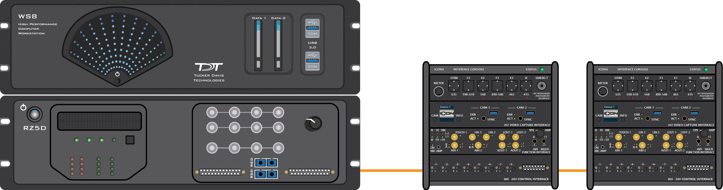
The iCon is a simple-to-use behavioral control interface designed to support easy or complex experiments. With interchangeable analog and digital modules to fit the needs of any experimental paradigm, it can control most third-party behavioral chambers.
The iCon connects directly to any TDT RZ processor. It can be controlled with TDT’s Synapse software — resulting in high-bandwidth, precision control of behavioral hardware with real-time signal monitoring. This connection allows for perfect synchronization between your behavioral tasks with your fiber photometry or electrophysiology data.
Research Uses
Product Specs
Resources
Publications
- Behavioral neuroscience
- Subject training
- Fiber photometry
- Electrophysiology
- Auditory research
- Available with two (iCon2) or four (iCon4) module slots
Publications
11 published studies used the iCon. The newest:
bioRxiv* · Sep 2026
Noise-induced hearing loss delays auditory learning and reduces the influence of acoustic evidence on choice (opens in a new tab)
Abstract
Noise-induced hearing loss (NIHL) degrades auditory sensitivity, but how it affects the learning of sound-guided decisions remains unclear. We trained adult Mongolian gerbils with normal-hearing (NH) or permanent NIHL, induced by a 2-hour exposure to 120 dB SPL broadband noise, to discriminate 4- versus 12-Hz amplitude-modulated (AM) broadband noise presented in a two-alternative forced-choice task. Stimuli were presented at comparable sensation levels across groups. Relative to NH animals, NIHL animals required approximately 2.5 times more sessions and 2.9 times more trials to reach task acquisition criterion. This learning delay was not explained by reduced task engagement, response latenc
eLife · May 2026
The nucleus accumbens shell regulates hedonic feeding via a rostral hotspot (opens in a new tab)
Abstract
The medial nucleus accumbens shell (medNAcSh) is a key regulator of hedonic feeding, controlling reward consumption through its projections to downstream structures. Recent studies showed that the primary cellular mediators of these effects are dopamine 1 receptor-positive striatal projection neurons (D1-SPNs). Specifically, D1-SPN activity gets inhibited during reward consumption, and such inhibition is necessary and sufficient to authorize consumption, independent of metabolic need. Anatomically, the medNAcSh spans 1–1.5 mm along the rostro-caudal axis in mice, and previous studies have reported functional gradients along this axis. For instance, pharmacological studies have suggested that rostral rather than caudal medNAcSh regulates appetitive behavior. However, the mechanisms underlying this topographical gradient remain unknown. Here, we hypothesized that D1-SPNs contribute to this gradient by regulating hedonic feeding via a specific hotspot in the rostral medNAcSh. Using calcium monitoring with fiber photometry in mice, we show that rostral medNAcSh D1-SPNs demonstrate inhibitory responses during reward consumption, while caudal D1-SPNs do not. Consistently, optogenetic stimulation of rostral D1-SPNs inhibits consumption, while stimulation of caudal D1-SPNs had minimal effects, confirming the existence of a functional rostro-caudal gradient. Importantly, we observed no differences between rostral and caudal D1-SPNs in their responses to aversive stimuli, suggesting that the D1-SPN gradient is specific to appetitive contexts. To investigate potential molecular correlates of this functional gradient, we leveraged open-source anatomy datasets and performed fluorescent in situ hybridization, identifying Stard5 and Peg10 as markers enriched in the rostral and caudal medNAcSh, respectively. Finally, we developed a novel Stard5-Flp driver line to selectively target the rostral hotspot and demonstrated that Stard5+ neurons recapitulate rostral D1-SPN activity patterns. Together, these findings establish a spatially confined rostral medNAcSh subregion as a critical regulator of reward consumption and introduce Stard5 as a molecular tool for its manipulation, offering new opportunities for intervention in dysregulated eating.
bioRxiv* · May 2026
Thalamus orchestrates local acetylcholine-dependent dopamine release in the learning striatum (opens in a new tab)
Abstract
Dopamine is essential for striatal function and learning. Striatal dopamine release can be triggered by dopamine cell firing, but also by coordinated cholinergic interneuron activity, which stimulates dopamine release via presynaptic nicotinic acetylcholine receptors on dopamine axons. While acetylcholine-dependent dopamine release is well-documented ex vivo and under artificial optogenetic stimulation in vivo , its role during natural behavior has remained unclear. One possible endogenous driver of acetylcholine-dependent dopamine release is thalamic input, which provides strong excitatory drive to cholinergic interneurons. To examine whether thalamic input provokes acetylcholine-dependent dopamine release during behavior, we performed simultaneous fiber photometry recordings of striatal dopamine (GRAB-rDA3m) and thalamic axon activity (gCaMP8m) in the dorsomedial (DMS) and dorsolateral striatum (DLS) of mice learning the accelerating rotarod, a striatal-dependent task that demands precise and effortful motor control. Recordings were obtained on- and off-task and across days of training to capture the full arc of learning. Dopamine transients in DMS, but not DLS, were frequently coupled to peaks in thalamic axon activity via an acetylcholine-dependent mechanism. The occurrence of these thalamic-evoked DMS dopamine transients depended on learning, task engagement, and the recent history of dopamine activity, but did not contribute to motor error signals. Together, these findings establish thalamic input as a physiological driver of acetylcholine-dependent dopamine release in DMS. Moreover, they reveal that striatal sensitivity to this local release mechanism is dynamically gated by dopaminergic history, providing a compelling framework for understanding how local and soma-triggered dopamine signals are coordinated to support learning.
* Preprint: not yet peer reviewed.
How to cite the iCon
Name the model in your Methods so readers can find the equipment you used, for example:
iCon RZ Interface Addition with Synapse software (Tucker-Davis Technologies, Alachua, FL, USA)
