Publication Spotlight: Greger Lab
December 21, 2022
Congratulations to Dr Guest and the Greger lab at Arizona State University for their recent publication in Clinical Neurophysiology. The paper is entitled “Microscale electrophysiological functional connectivity in human cortico-basal ganglia network”. DOI: 10.1016/j.clinph.2022.06.017
Significance:
Deep Brain Stimulation (DBS) is a therapeutic tool used to treat motor disabilities in disorders such as Parkinson’s Disease (PD). However, not much is known about the specific mechanism through which DBS alters the electromagnetic field within the cortico-basal ganglia network to improve motor coordination. This paper aims to investigate the electrophysiological relationships in the cortico-basal ganglia circuit during DBS to better understand the functional relationships of the brain regions in a PD model.
Methodology:
During DBS electrode placement, Guest et al. performed human in vivo electrophysiological recordings of PD patients from the subthalamic nucleus (STN) – using microelectrodes – and the motor association cortex – using a micro-electrocorticography grid. STN action potential recordings and motor association cortex local field potentials (LFPs) were measured to determine functional connectivity between the subcortical and cortical regions.
Results:
Guest et al. found a subset of neurons in the STN that exhibited functional connectivity with the LFPs of the STN, as well as the LFPs in the motor association cortex. This spatial topography exists on a sub-centimeter scale, indicating a high spatial specificity. While some STN neurons exhibited high functional connectivity with the STN LFPs and the cortical LFPs, other neurons from a few hundred microns away showed no functional connectivity. This indicates that within larger anatomical connections of the cortico-basal ganglia network, there are subsets of smaller, micro-scale circuits involving subsets of neurons, rather than a uniform connection of neurons between brain regions. This study also demonstrates the utility of micro-ECoG devices for electrophysiological recordings in research settings.
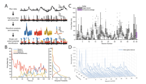
A.) Illustration of the workflow for isolating action potentials and spike sorting. B.) Firing rate of 3 example STN neurons during the same recording. C.) Firing rates of all 46 STN neurons recorded. D.) Inter-spike intervals of all 46 STN neurons.
TDT Equipment Used:
Electrophysiological recordings were performed with processors and equipment from Tucker-Davis Technologies. TDT offers systems for both lower channel count recordings (up to 32 channels) with the RZ5D processor and higher channel count recordings (up to 256 channels) with the RZ2 base processor. Guest et al. used the TDT data acquisition system for continuous 16-channel recordings of the STN and motor association cortex to determine the functional connectivity between the two regions.
Current Lab Focus:
The Greger Lab continues to use in-vivo microelectrode arrays to study neural function and pathology. By comparing action potentials to local field potentials in different cortical regions, the lab has been able to decode certain behaviors – such as spoken words and finger movements – from neural signals that have been recorded on these microelectrode arrays. As evidenced by this recent publication, the lab is currently using this approach to better understand connectivity in the cortico-basal ganglia network in disease models such as Parkinson’s Disease.