Fiber Photometry User Guide
Hello and welcome to the Fiber Photometry User Guide. We appreciate you taking the time to view this document. First, if you are a TDT customer, then thank you - we greatly appreciate your business and we hope to help you meet your research goals. If you are considering purchasing a fiber photometry system from us, then thank you as well - TDT is the industry leader in fiber photometry systems and we have many successful and happy customers who use our products. We would enjoy nothing more than to have you join the TDT family.
Tip
We recommend you visit the main TDT Fiber Photometry page on our website to learn more about purchasing a new system.
The objective of this document is to be a hardware and software instructional reference for all levels of fiber photometry users. This guide will not go into any meaningful details about the biological underpinnings for fiber photometry, calcium (Ca++) imaging, optogenetics, or other related fields. The successful use of your fiber photometry equipment is predicated on you knowing how to get fluorophores to express in cells and perform surgeries for in vivo monitoring of neural targets.
Definitions
This section includes brief definitions for keywords you will read throughout the guide.
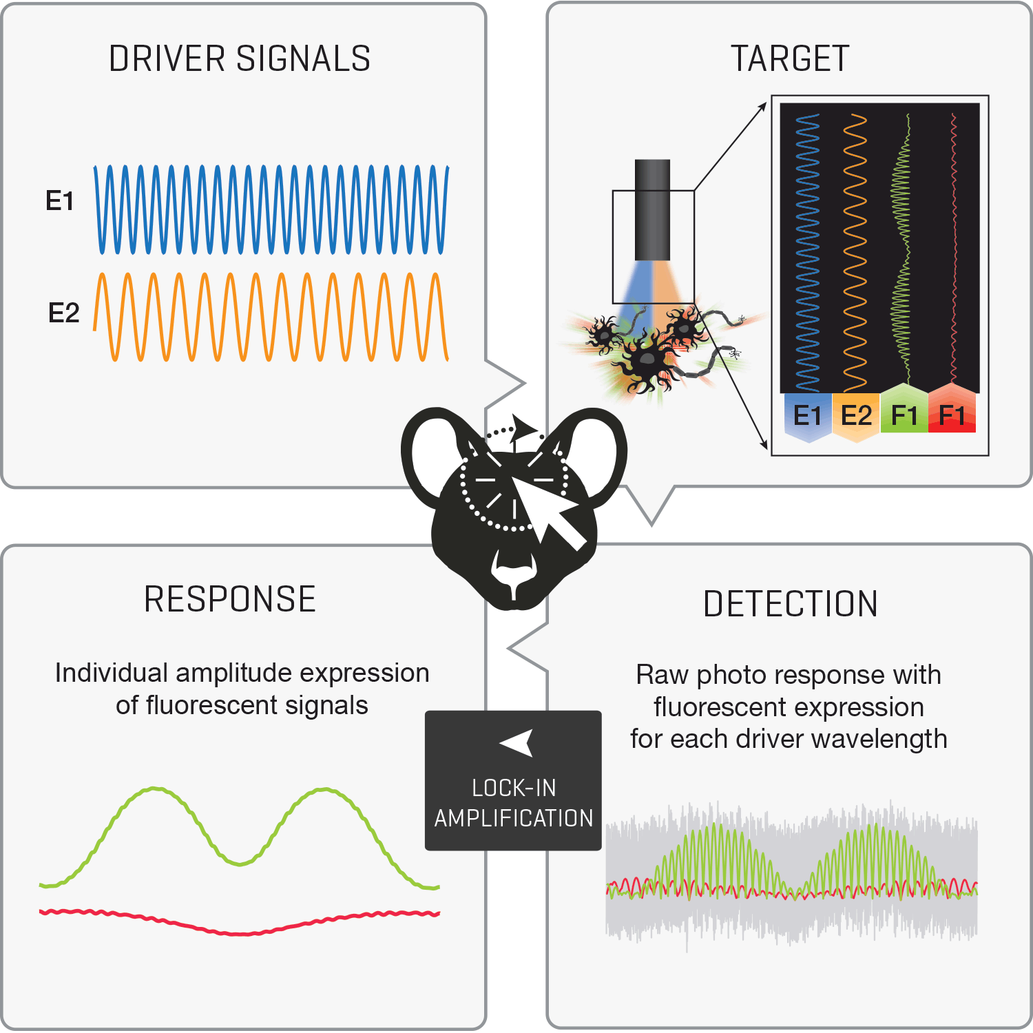
Fiber Photometry: An imaging technique used to monitor neural activity of specifically-targeted cell populations. Fiber photometry uses excitation light from implanted fiber optics to record fluorescent activity of genetically-encoded calcium indicators (GECI) in neuronal populations.
GCaMP: GCaMP is a GECI that fluoresces in the presence of calcium (Ca++) activity in neurons. For more about GCaMP please see Janelia's definition page https://www.janelia.org/open-science/gcamp.
Isosbestic: This is your control signal that will be used to correct for motion artifact and photobleaching in post-processing.
"In spectroscopy, an isosbestic point is a specific wavelength, wavenumber or frequency at which the total absorbance of a sample does not change during a chemical reaction or a physical change of the sample." Wikipedia
GFP: Green Fluorescent Protein. This is the protein coupled into GCaMP that fluoresces at a 510 nm peak when excited by a peak 488 nm light source https://www.fpbase.org/protein/egfp/.
Autofluorescence: The emission of light from either fiber optic components or brain tissue when excitation light is absorbed. Autofluorescence (AF) is parasitic and increase the overall background noise in recordings; removing AF as much as possible via using low AF subject cables and photobleaching patch cords is important.
Photobleaching (GFP): The overexposure of GFP to a light source that involves an irreversible change in the structure of the GFP protein. Long-term low-level light exposure and high- intensity light exposure will cause photobleaching. With photobleaching, users will see a decrease in response from the GFP and the response will be at a constant lower level.
Photobleaching (Patch Cords): The process of exposing a fiber optic patch cable to high levels of light (500 mA) for a long duration (~4 hours) to reduce auto fluorescent emissions from within the cable.
Demodulated: The demodulated signals are your response waveforms. These are the relevant fluorescence data that have been extracted from the raw photosensor signal and low pass filtered using lock-in amplification. You should think of these data as being close to an un-normalized and corrected dF/F or z-score.
Lock-in Amplification: Lock-in amplification is a signal processing technique that uses modulation of driver signals and an orthogonal reference signal to extract relevant amplitude and phase of frequency-specific responses in a complex and often noisy signal. Please see the following diagram.
dF/F and z-score: Mathematical paradigms used to normalize and quantify relative change of a continuous time series. These are commonly used metrics in the calcium imaging field.
Important LED Safety Information
Caution must be used when operating the LUX LEDs. High power light output from the LUX LEDs can be harmful to the eyes and skin. Never look directly at any LED light output, either from the LED module directly or from the output of a connected optical cable. For ultra-violet (UV) LEDs (415 nm and lower), extra precaution must also be taken to avoid direct light exposure to skin. Protective eyewear, such as these from ThorLabs, should be worn when operating LEDs.